June 4, 2025
Neighbourly help in the brain: Nerve cells step in when lost
A previously unknown mechanism allows networks in the brain to reorganize themselves after damage.
How the brain largely maintains its function when neurons are lost – this is what researchers at the Mainz University Medical Center, the Frankfurt Institute for Advanced Studies (FIAS) and Hebrew University (Jerusalem) have deciphered. They show that neuronal networks in the cerebral cortex reorganize within a short period of time, with other nerve cells taking over the tasks of the lost neurons. These findings could form the basis for future research into natural ageing processes and neurodegenerative diseases such as Alzheimer's or Parkinson's. The study was published in the renowned journal Nature Neuroscience.
Nerve cells (neurons) are the most important building blocks of the brain. They form the basis for all mental and physical functions such as thinking, feeling, movement, and perception. In the course of life, nerve cells in the brain can be lost for various reasons: They die off due to age-related processes, are damaged by toxins such as alcohol, or neurodegenerative diseases such as Alzheimer's and Parkinson's lead to a more rapid progressive loss of neurons.
While most body organs regularly replace old or damaged cells with new ones in order to maintain their organ function, new neurons only form in certain regions of the brain. In the cerebral cortex, which is responsible for complex thought processes and perception, the ability to form new neurons is very limited in adulthood. “Nevertheless, clinical studies have shown that cortical brain function is often surprisingly resistant to the loss of neurons that occurs in the course of aging or neurodegenerative diseases,” explains Simon Rumpel, head of the Systems Neurophysiology research group at the Institute of Physiology at the Mainz University Medical Center.
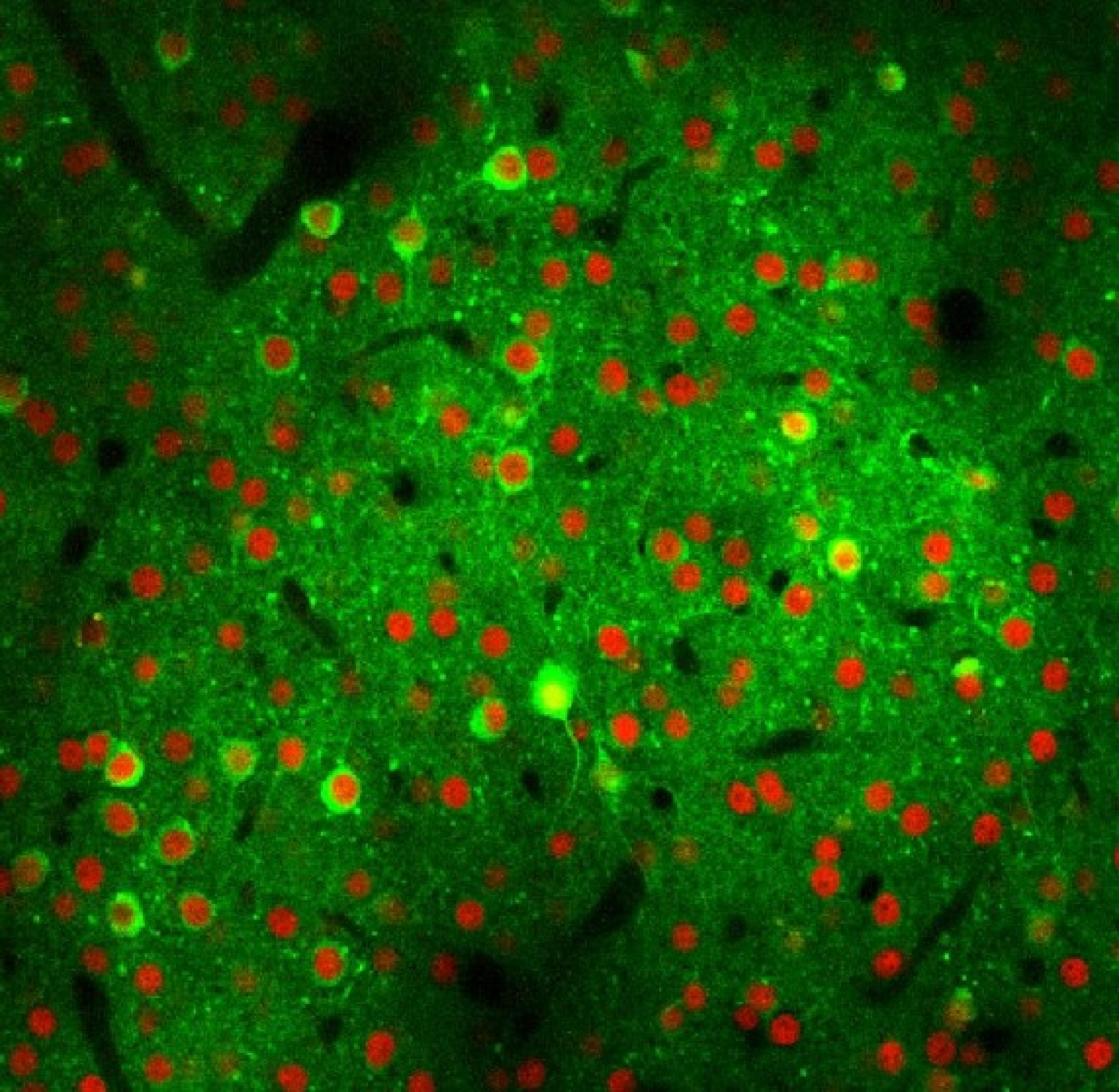
Until now, it was not known how the brain can compensate for the loss of nerve cells and maintain its function. To find this out, the research team used an animal model to investigate the neuronal networks in the auditory cortex, which is responsible for processing acoustic stimuli. The perception of sounds is based on activity patterns that are triggered in the brain by acoustic stimuli. These patterns are very complex. PhD student Bastian Eppler and Senior Fellow Matthias Kaschube at FIAS contributed significantly to the analysis of these data and the interpretation of the results with their expertise.
The researchers found that the activity patterns initially destabilize when the loss of a few specific nerve cells is deliberately induced. This indicates that the neuronal network responsible for sound perception is in a delicate balance. After just a few days, very similar activity patterns form again. The nerve cells that were not previously activated by the acoustic stimuli now acquire the ability to take the place of the lost neurons. “We assume that this newly discovered neuronal mechanism plays an important role in the loss of nerve cells in natural ageing processes as well as in neurodegenerative diseases,” says Rumpel. Future research efforts could aim to support this neuronal reorganization.
Publication: Takahiro Noda, Eike Kienle, Jens-Bastian Eppler, Dominik F. Aschauer, Matthias Kaschube, Yonatan Loewenstein, Simon Rumpel: Homeostasis of a representational map in the neocortex; Nature Neuroscience (2025), DOI: 10.1038/s41593-025-01982-7